

Who Was The First To Discover That Exposure To X-rays Could Produce Burns To The Skin?
The X-Ray Era: 1901–1915
Dr. Pusey'southward Radiation Handling for Breast Cancer, Chicago: 1901

The text and photographs on this page are excerpted from a four-volume serial of books titled Oncology: Tumors & Handling, A Photographic History, by Stanley B. Burns, MD, FACS, and Elizabeth A. Burns. The photos below are from the volume titled "The 10-Ray Era: 1901–1915." The photographs appear courtesy of Stanley B. Burns, Physician, and The Burns Archive. To view boosted photos from this series of books, visit burnsarchive.com.
Inside 2 months of its discovery, x-ray's invisible light was being used to care for breast cancer. Chicago medical student Emil H. Grubbe was making Crookes vacuum tubes for physicists when Wilhelm Röntgen announced his discovery of the x-ray in November 1895. Mr. Grubbe immediately began modifying the tubes to produce ten-rays. He noticed his hands developed burns while working with the new tubes, and a professor at the medical school hypothesized that they were acquired by the new rays. Hoping to "burn down out" cancer, on January 29, 1896, one of Mr. Grubbe'due south professors allowed him to use x-rays to treat a patient who had a recurrent breast carcinoma. With the credible success of the treatment, 17 more sessions were carried out, and radiation therapy was born.
In 1893, prior to Mr. Röntgen's discovery of the x-ray, Dr. Niels Ryberg Finsen (1860–1904) set up an important new therapeutic precedent when he used the curative power of ultraviolet rays to treat peel diseases. Dr. Finsen'south discovery, chosen photo or light therapy, won him the 1903 Nobel Prize. During the starting time ii decades of the 20th century, both x-ray and phototherapy were developed and became important therapeutic modalities. Phototherapy effectively treated and cured not only pare diseases, merely tuberculosis, the primary killer in this era, every bit well. Tens of thousands of patients were saved past its use, yet Dr. Finsen's name and accomplishments are relatively unrecognized today.
By 1903, several texts were produced extolling the virtues of 10-rays. In July 1903, William Allen Pusey, Md, Professor of Dermatology at the University of Illinois, and Eugene Wilson Caldwell, an engineer and Director of the Gibbs X-ray Laboratory University and Bellevue Hospital in New York, published a 670+ page text, The Practical Application of the Roentgen Rays in Therapeutics and Diagnosis. In this landmark work, produced only 7 years after the discovery, Dr. Pusey outlined his techniques for treating dermatitis; tuberculosis; cutaneous carcinoma; chest cancer; and deep-seated cancers of the esophagus, abdomen, rectum, and larynx likewise as sarcoma and leukemia. Dr. Pusey's emphasis was on the "prophylactic use" of radiation, significant its employ afterward surgery to foreclose recurrence.
Radiation Therapy for Recurrent Breast Cancer
The use of radiation to treat recurrent carcinoma of the breast is documented in these photographs. The first photograph was taken on Nov 1, 1901, and the 2d was taken on February 25, 1902. His example written report reads as follows:
Mrs. X, aged 58, referred to me by Dr. C.C. Gratiot of Shullsburg, Wisconsin. (The patient was in the care of Christian Fenger, MD, in Passavant Infirmary in July and August 1900.) Her nowadays (July 1900) trouble began most 6 years ago, when the patient noticed a little hard lump on the inferior aspect of the left breast on the axillary side. There was scarcely perceptible enlargement of the hard area until about 2 years ago, when the patient noticed a more rapid enlargement. She then consulted Dr. Sheldon, of Madison, Wisconsin. At that fourth dimension, the growth developed very fast and broke through the skin and formed an open sore…, which grew in dimensions until it is now the size of a silver dollar.
Dr. Sheldon brash an operation, whereupon she consulted Dr. Fenger. Dr. Fenger removed the breast with the axillary and supraclavicular glands in July 1900. A few months after [its] removal, the affliction reappeared in the scar. She returned to Dr. Fenger final summer, but he found the case across operation. Her condition when she came to me, November ane, 1901, is shown in the accompanying photograph [Figure 1]…. There was a mass of carcinomatous tissue on the left side involving an area about a square foot. The nodules varied from the size of a hazelnut to that of a small apple tree. At the upper angle there were a couple of ulcers 2.5 to 3 inches in diameter….
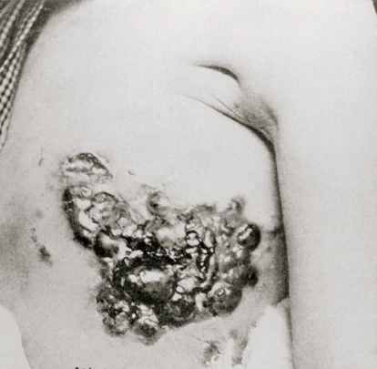
Effigy i: Pretreatment photo shows a mass of carcinomatous tissue.
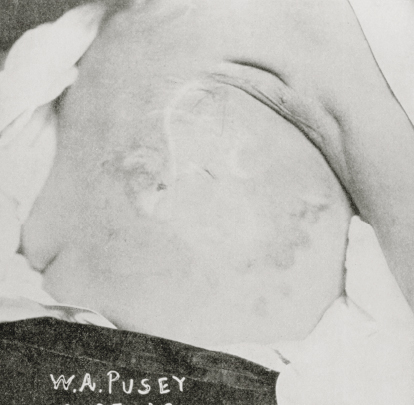
FIGURE ii: Posttreatment photo shows no bear witness of carcinoma on the chest wall.
When she began handling, she was suffering a slap-up bargain of pain… [and] was haggard and worn…. She was put under ten-ray exposures on Nov one, and the sittings were given daily for two months and every other twenty-four hour period or less ofttimes after that. 2 weeks after the treatments were begun…, her pain was gone, and she has had none since.
On January 17: There had been rapid subsidence of the tumor the last calendar month…, There is at present but a slight superficial ulceration…and one pocket-sized nodule…. The surface is gratis from infiltration and thickening and the redness has almost disappeared. The peel is considerably pigmented…. Comeback has continued since that fourth dimension. The condition on Feb 27, 1902, is indicated…[in Figure 2]. In that location is no evidence of carcinoma left on the breast wall…. The first series of treatment was ended….
During May and June 1902, she had rather vigorous exposures, with the production of an astute dermatitis, which disappeared slowly…. From September to October she had vigorous exposures… [and] the lesions disappeared in all of the locations…. December 15, 1902: She remains in skillful status without evidence of carcinoma.
[Dr. Pusey commented that this patient would have died near the fourth dimension of her outset treatment in November 1901.] This case tends to disprove the idea… hat afterwards a sure amount of exposure carcinomatous tissue is no longer susceptible to the influence of x-rays…. In the spring of 1903 the affliction recurred…involvement of the lung appeared, and the patient died [in] October 1903, refusing information technology, on the ground that, her children died during the preceding year, [and] she had no desire to brand whatsoever farther effort to prolong her life. ■

Source: https://ascopost.com/issues/june-10-2018/the-x-ray-era/
Posted by: stoutonsing.blogspot.com
0 Response to "Who Was The First To Discover That Exposure To X-rays Could Produce Burns To The Skin?"
Post a Comment